Questions about a product, technology or experimental design? Give us a call 610-644-8845.

VU101: Anti-Ubiquitin Antibody (MAb; clone VU-1)

LifeSensors VU101 is a mouse monoclonal anti-ubiquitin antibody (clone VU-1) that detects poly- and monoubiquitylated proteins and free ubiquitin, supporting ubiquitin pathway research.

$499.00$939.00

SKU VU-0101 Categories ,
  • Imunnohistochemical staining: 1:200 – 1:500
  • Western blots: 1:1000 – 1:10,000 — Please note: To obtain maximum sensitivity, the membrane should be treated with 0.5% glutaraldehyde prior to exposure to the primary antibody
  • Immunohistochemistry using chromogenic substrates with a low backgound (see information on the Datasheet);
  • Immunofluorescence in tissue sections and cell culture;
  • Western blot analysis to detect mono- and polyubiquitinated proteins as well as free ubiquitin
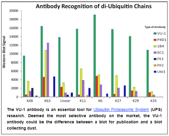
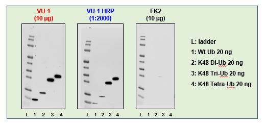
LifeSensors has developed several monoclonal antibodies that recognize polyubiquitylated proteins and free ubiquitin. VU-1 has been shown to recognize all ubiquitin linkages (mono, K6, K11, K27, K29, K33, K48, K63, and linear) by Western blotting. VU-1 is an excellent antibody for immunostaining applications generating robust ubiquitin detection and low background with an ability to detect subcellular ubiquitin localization. Ubiquitin (Ub) is a highly conserved, 8 kDa polypeptide present in all eukaryotic cells. Ubiquitin is conjugated to the ε-amino group of lysine residues in the target protein through the sequential action of a three-tiered enzymatic pathway.  The following are the three enzymes: an E1 Ub activating enzyme, an E2 conjugating enzyme, and an E3 ligase. In addition, the seven lysines within Ub itself can serve as Ub acceptors leading to the formation of polyubiquitin chains. Among these K48 and K63 linkages are well characterized. Specifically, K48-linked polyubiquitylation targets proteins for proteasomal degradation whereas K63 linkages regulate signaling events, receptor endocytosis and immune responses. However, polyubiquitin linkages at other lysines are less prominent and their physiological role remains largely unknown.

Info

Host Mouse
Quantity 50 µg, 250 µg
Concentration Variable
Storage -20°C; Avoid freeze/thaw cycles
  Product ships with 1 vial of Glutaraldehyde.
  • Imunnohistochemical staining: 1:200 – 1:500
  • Western blots: 1:1000 – 1:10,000 — Please note: To obtain maximum sensitivity, the membrane should be treated with 0.5% glutaraldehyde prior to exposure to the primary antibody
  • Immunohistochemistry using chromogenic substrates with a low backgound (see information on the Datasheet);
  • Immunofluorescence in tissue sections and cell culture;
  • Western blot analysis to detect mono- and polyubiquitinated proteins as well as free ubiquitin

send us a message for pricing

CONTACT INFORMATION

Phone (Office)

610.644.8845

SEND EMAIL

info@lifesensors.com

VISIT ANYTIME

271 Great Valley Parkway Malvern, PA 19355

More from This Category

PROTAC IS A REGISTERED TRADEMARK OF ARVINAS OPERATIONS, INC., AND IS USED UNDER LICENSE.